
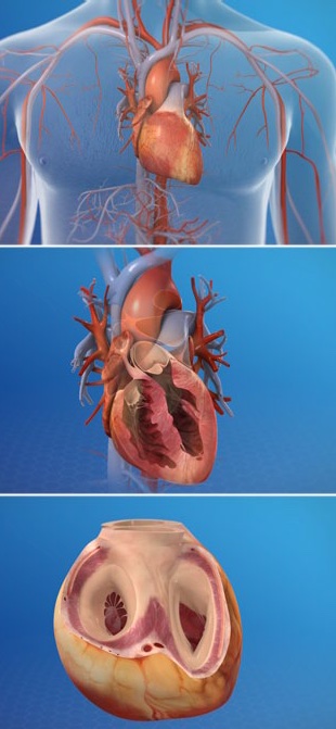
The heart is a muscular organ that pumps blood throughout your body.
It is positioned behind the lungs, slightly to the left side of the chest.
Your heart is a bit larger than the size of your fist.
The Right Side
The heart is divided into two sides and four chambers. On the right side, blood that has already circulated through the body enters the heart through the superior vena cava and the inferior vena cava. The blood flows into the right atrium. When this chamber is full, the heart pushes the blood through the tricuspid valve and into the the next chamber – the right ventricle. From there, the blood is pushed out of the heart through the pulmonary valve. The blood travels through the pulmonary artery to the lungs, where it will pick up oxygen and give up carbon dioxide.
The Left Side
On the left side of the heart, blood that has received oxygen from the lungs enters the heart through the pulmonary veins. The blood flows into the left atrium. It is pushed through the mitral valve into the left ventricle. Finally, it is pushed through the aortic valve and into the aorta. The aorta is the body’s largest artery. It helps distribute the oxygen-rich blood throughout the body.
The Valves
Now let’s take a closer look at the valves. All of the heart’s valves allow blood to flow one way but not the other. They open to let blood pass through, and then quickly snap shut. They keep blood moving in the right direction through the heart. The tricuspid and mitral valves are called the “atrioventricular” valves. The tricuspid valve has three flaps (called “cusps” or “leaflets”) and the bicuspid valve has two. These valves are connected to the papillary muscles of the heart by thin, tendon-like strings called chordae tendineae. The pulmonary valve and the aortic valve are called the “semilunar” valves. They normally have three flaps.
The Heartbeat
Now let’s examine the heartbeat. The heartbeat is triggered by an electrical impulse. This impulse is generated by the sinoatrial node, a specialized group of cells in the wall of the right atrium. The impulse causes the upper chambers to contract. Then, it travels to the lower chambers, causing them to contract and to push blood out of the heart.
Coronary Arteries and Veins
Now let’s look at the heart’s exterior. On the exterior of the heart are the coronary arteries and cardiac veins. The arteries supply the heart muscles with oxygen-rich blood. The veins carry away blood when the oxygen and nutrients have been depleted. In addition to these vessels, pads of fatty tissue are also found on the exterior of the heart. And finally, the entire heart is wrapped in a double-layered sac called the “pericardium.”
Caring for the Heart
The heart is a complex and hardworking organ. It is susceptible to cardiovascular disease, which can lead to heart attack and stroke. Your diet, your lifestyle and your activity level can all have an impact on your heart’s health.